Free images, micrographs, and drawings of Acinetobacter baumannii (colonies, cells, genomes, genetic elements, etc.)


Colonies of A. baumannii strain AB5075 showing phase vaiation
Photo Credit: Nabil Karah, M.d., Ph.D., Department of Molecular Biology, Umeå University
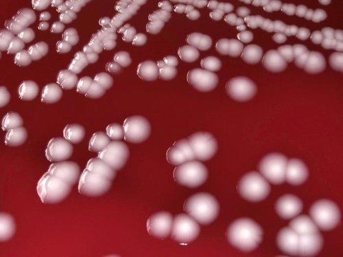
Photo Credit: Todd Parker, Ph.D., Assoc Director for Laboratory Science, Div of Preparedness and Emerging Infections at CDC.
Under the low-power magnification of 10X, using a digital Keyence scope, this photograph depicts the colonial growth displayed by the Gram-negative bacterium, Acinetobacter baumannii, which were cultured on sheep blood agar (SBA) medium, for a 48-hour time period, at a temperature of 37°C.
Creator: CDC/ Matthew J. Arduino, DRPH; Janice Carr; Jana Swenson
Source: Public Health Image Library
http://www.publicdomainfiles.com/show_file.php?id=13522305613715
All the images are free of copyright restrictions.
Would you like to share your images of A. baumannii, just send us an email.
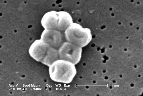
This SEM depicts a highly magnified cluster of Gram-negative, non-motile Acinetobacter baumannii bacteria; Mag - 27600x.